





Retinal detachment is an eye condition that should be examined by an ophthalmologist as soon as possible, especially retinal detachment caused by a retinal tear, which often requires surgical treatment. If left untreated until it involves the macula (the central area for sharp vision), it may cause permanent loss of vision.
What is the retina?
The retina (Retina) is a thin layer of tissue lining the back inside of the eyeball, consisting of millions of photoreceptor cells and other nerve cells. It receives light and sends signals to the brain, allowing us to see images. If an abnormality occurs, it can reduce visual clarity.
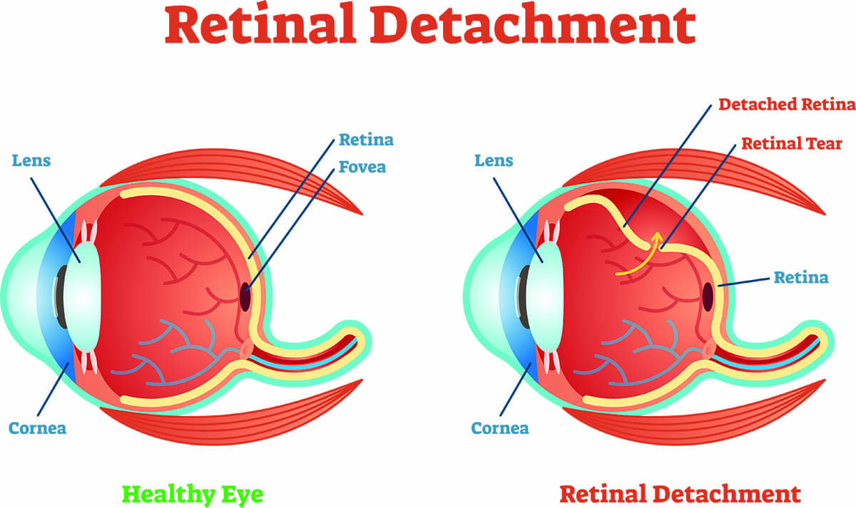
What is retinal detachment?
Retinal detachment (Retinal Detachment) is a condition in which the retinal layer, a thin tissue lining the inside of the eyeball, separates from the tissue and blood vessel layer behind it that nourishes the retina. This causes the photoreceptor cells in the detached area to function abnormally, and if left untreated for a prolonged period, it may result in permanent damage, making it impossible for vision to return to normal.
What causes retinal detachment?
- Vitreous degeneration that pulls on the retina until it tears (Rhegmatogenous Retinal Detachment)
- Traction from fibrous tissue inside the eye, such as in patients with advanced diabetic retinopathy
- Fluid accumulation under the retina due to inflammation or an intraocular tumor
- Accidents or severe blunt trauma to the eye
Risk factors for retinal detachment
- Severe myopia
- Previous cataract surgery
- A history of retinal detachment in the other eye or a family history of retinal detachment
- Previous eye injury

What are the symptoms of retinal detachment?
- Early stage
- Detachment limited to the peripheral edge of the retina and not yet involving the macula
- Seeing lightning-like flashes or flash lights (Flashing) in one or both eyes
- A sudden increase in black spots or black lines (Floaters) in the vision
- Progressive stage
- The detachment extends closer to the macula
- A curtain or dark shadow partially blocks one side; central vision remains clear, with a sensation that half of the image is missing
- Macular detachment stage
- The detachment has reached the macula
- Sudden blurred vision; central vision becomes unclear or disappears
How is retinal detachment diagnosed?
If you have abnormal symptoms, you should see an ophthalmologist for a detailed eye examination. After dilating the pupils with eye drops, the ophthalmologist will use special instruments and lenses to examine the retina all around. If no retinal tear is found and there is no retinal detachment, the doctor will schedule another visit within 1 – 2 weeks for a repeat examination to confirm that there is truly no tear or detachment. After pupil dilation, you may have blurred vision and light sensitivity for about 4 – 6 hours until the pupils return to normal. After that, if new abnormal symptoms occur or symptoms worsen, you should see an ophthalmologist immediately. If the ophthalmologist cannot view the retina with standard instruments, such as when there is vitreous hemorrhage, an ophthalmic ultrasound (Ophthalmic Ultrasound) may be needed to help diagnose retinal detachment.
Why retinal detachment must be treated urgently
When the retina separates from the nourishing tissue layer, photoreceptor cells function abnormally, and if left for a long time, permanent damage may occur. The closer the detachment is to the macula or once it involves the macula (Macula), and the longer it is left untreated, the lower the chance that vision will recover well after treatment. Therefore, if retinal detachment is suspected, you should be examined by an ophthalmologist as soon as possible to assess the location and severity of the detachment and plan appropriate treatment, and you should not wait for vision to become more blurred.

Approaches to treating retinal detachment
- Laser photocoagulation alone (Laser Photocoagulation) may be used only in patients with a small retinal detachment at the far peripheral edge of the retina. If the detachment is extensive, laser treatment alone is not sufficient.
- Injection of a gas bubble into the eye together with laser treatment or cryotherapy (Pneumatic Retinopexy) around the retinal tear to prevent fluid from passing through the tear. Hospitalization is not required. After treatment, avoid activities that may involve impact or trauma to the eye for about 1 – 2 weeks to allow firm adhesion to form around the tear before healing. This method is suitable only in certain cases.
How is retinal detachment surgery performed?
Retinal detachment often requires surgical treatment, which is divided into 2 methods:
- Scleral buckling using a silicone band or sponge placed on the outside of the eyeball (Scleral Buckle)
- Pars plana vitrectomy and direct internal retinal repair (Pars Plana Vitrectomy – PPV), which is commonly used today, to remove the vitreous and any tractional membranes pulling on the retina (if present), reattach the retina, and apply laser treatment or cryotherapy as appropriate. A gas bubble may then be used to help press the retina back into place. The patient may need to maintain a face-down position or keep the head in a specific position as determined by the doctor; the duration depends on the location of the detachment, the surgical method, and the type of gas used. In some cases, silicone oil (Silicone Oil) may be chosen instead of gas, and a later surgery may be required to remove the silicone oil after the retina has reattached well.

Postoperative care after retinal detachment surgery
- If surgery uses an intraocular gas bubble, the patient may need to maintain a face-down position or hold the head position strictly as advised by the doctor. The duration depends on the location of the detached retina, the surgical method, and the type of gas used. Do not travel by airplane or go to high-altitude areas with low air pressure until the gas in the eye is fully absorbed, because the gas may expand and cause an acute rise in intraocular pressure, leading to severe eye pain and potential harm to vision.
- After surgery, you may temporarily notice floating black spots. If an intraocular gas bubble was used, you will see the shadow of the gas bubble, which will gradually lower as the gas is absorbed on its own. How long the gas remains in the eye depends on the type and amount of gas used and may take several weeks.
- The operated eye may feel irritated or uncomfortable. Do not rub the eye. Artificial tears may be used to relieve irritation. If there is pain, pain relievers may be taken. If symptoms are severe, see an ophthalmologist.
- During the first week after surgery, the white of the eye (conjunctiva) may be swollen and red, and the eyelid may be swollen. This is often inflammation or swelling from face-down positioning after surgery. Use eye drops as prescribed by the doctor. If there is a lot of eye discharge, severe eye pain, or worsening vision, see an ophthalmologist urgently.
- Wear an eye shield for 2 – 4 weeks or as directed by the doctor. During the day, glasses or sunglasses may be used.
- Avoid getting water into the operated eye for about 2 – 4 weeks after surgery to prevent infection inside the eye.
- Avoid forceful coughing, sneezing, or straining, as it may cause bleeding in the eye.
- Rest and refrain from exercise or activities that may impact or traumatize the eye.
- Vision will not improve immediately after surgery. If the surgery is successful and the retina reattaches well, vision may
gradually improve over several weeks after surgery. In some cases, recovery may take as long as 2 – 3 months or longer, depending on the severity and location of the detachment.
How to prevent retinal detachment
- See an ophthalmologist for an eye and retinal examination at least once a year, especially for people with severe myopia or other risk factors.
- Prevent eye injuries by wearing protective eyewear (Safety Goggles) when doing high-risk activities or playing certain sports.
- If abnormal symptoms occur, see an ophthalmologist as soon as possible.
Frequently asked questions
Q: What is the difference between retinal detachment and vitreous degeneration?
A: Vitreous degeneration is an age-related change of the vitreous gel, while retinal detachment is a condition in which the retinal layer separates from the wall of the eye. Vitreous degeneration can cause retinal detachment in some people, but not everyone with vitreous degeneration will develop retinal detachment.
Q: After retinal detachment surgery, will vision return to normal?
A: If surgery is performed early before the detachment reaches the macula, the chances of visual recovery are generally good. However, if the detachment has already involved the macula, even if surgery is successful, central vision may not return to its previous sharpness.
Q: Can retinal detachment recur?
A: Retinal detachment can recur, especially in the same eye or the other eye in people with risk factors such as severe myopia. Therefore, regular follow-up with an ophthalmologist is recommended even after successful surgery.
Hospitals specializing in retinal detachment treatment
The Eye Center, Bangkok Hospital, is ready to provide care and treatment for retinal detachment with specialized ophthalmologists with expertise, modern tools and equipment, and an experienced multidisciplinary team. They are ready to plan care, treatment, and provide close consultation—from diagnosis and treatment through follow-up—to ensure confident vision at every age.
Physicians specializing in retinal detachment care and treatment
Dr. Weeraya Pimolrat Retina and vitreous specialist and ocular immunology specialist, Bangkok Hospital
You can click here to make an appointment yourself.