What is Cardiac MRI?
Magnetic resonance imaging (MRI) is a noninvasive medical test that uses a powerful magnetic field, radio frequency pulses and a computer to produce detailed pictures of organs, soft tissues, bone and virtually all other internal body structures. MRI does not use ionizing radiation (x-rays).
What does MRI scanner look like
Outside
The MRI scanner is on the left. The CT scanner is on the right. They look like giant donut from the front. The MRI scanner is usually deeper, although there are some variations such as short-bore systems, larger diameter bore and open on the sides (open MRI) which is also available at Bangkok heart Hospital.
Inside
The MRI scanner has powerful magnet and radio frequency coil while the CT scanner has X-rays tube at 2 o’clock and CT detectors are on opposite side at 8 o’clock. Only the CT scanner that spins around the patient at very fast spee
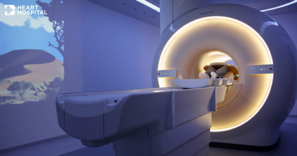
How does the MRI scanner work?
Unlike conventional x-ray examinations and computed tomography (CT) scans, MRI does not depend on ionizing radiation. Instead, while in the magnet field, radio waves redirect alignment of hydrogen atoms that naturally exist within the body without causing any chemical changes in the tissues. As the hydrogen atoms return to their usual alignment, they emit energy that varies according to the type of body tissue in which they lie. The MR scanner picks up this energy and creates a picture of the tissues scanned.
The magnetic field is produced by passing an electric current through wire coils in most MRI units. Other coils may be placed around the part of the body being imaged, send and receive radio waves, producing signals that are detected by the coils.
A computer then processes the signals and generates a series of images, each of which shows a thin slice of the body. The images can then be studied from different angles by the interpreting radiologist. Frequently, the differentiation of abnormal (diseased) tissue from normal tissues is better with MRI than with other imaging modalities such as x-ray, CT and ultrasound.
What are some common uses of the procedure? (Indication)
Cardiac MRI imaging is performed to help your physician detect or monitor cardiac disease by:
- Evaluating the anatomy and function of the heart chambers, valves, size and blood flow through major vessels, and surrounding structures such as the pericardium (the sac that surrounds the heart).
- Diagnosing a variety of cardiovascular (heart and/or blood vessel) disorders.
- Evaluating the effects of coronary artery disease such as limited blood flow to the heart muscle and scarring within the heart muscle after a heart attack.
- Planning a patient’s treatment for cardiovascular disorders.
- Monitoring the progression of certain disorders over time.
- Evaluating the anatomy of the heart and blood vessels in children and adults with congenital heart disease. Evaluating the effects of surgical changes, especially in patients with congenital heart disease.
How is the procedure performed?
MRI examinations may be performed on outpatients or inpatients.
- You will be positioned on the moveable examination table. Straps and bolsters may be used to help you stay still and maintain the correct position during imaging.
- You will be attached with electrocardiogram (ECG) leads, a respiratory gating belt, a small pulse monitor may be placed on your finger.
- You will be given breathing instructions and will be asked to hold your breath numerous times during the examinations. If a contrast material will be used in the MRI exam, there will be an intravenous line (IV). The contrast material will be injected into the IV line the after an initial series of scans. Additional series of images will be taken during or following the injection.
How long does the procedure take?
MRI exams generally include multiple runs (sequences), some of which may last several minutes. The entire examination is usually completed in less than 90 minutes once imaging has started, but may be shorter or longer depending on what the initial image shows.