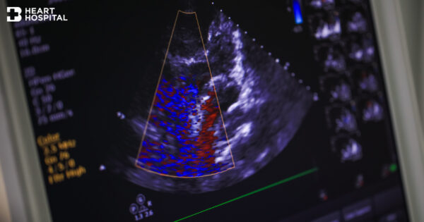
What is a CTA or Coronary CTA?
Computed Tomographic Angiography (CTA) is an imaging exam that uses a CAT (Computerized Axial Tomography) scanner to examine the cardiovascular system (heart and blood vessels in various parts of the body). After intravenous contrast injection in an arm vein, this highly specialized procedure utilizes x-rays to acquire axial images, which are then assembled into three-dimensional views of the blood vessels using state-of-the-art computer software. When used to view the arteries of the heart, this exam is referred to as coronary CTA.
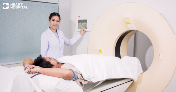
A CT scanner looks like a big doughnut. The patient aperture (opening) is 60 cm to 70 cm (24″ to 28″) in diameter. Inside the covers of the CT scanner is a rotating frame which has an x-ray tube mounted on one side and the detector mounted on the opposite side. A fan beam of x-ray (see the above figure) is created as the rotating frame spins the x-ray tube and detector around the patient (see figure below). Each time the x-ray tube and detector make a 360° rotation, an image or “slice” has been acquired. This “slice” is collimated (focused) to a thickness as thin as 1 mm