Finding a new mole or noticing that an existing spot has changed can be worrying. Fortunately, most moles are benign, but certain changes deserve assessment by a dermatologist—particularly changes in size, shape, color, or symptoms such as persistent bleeding or crusting.
A mole check involves more than simply deciding whether a spot “looks normal.” A dermatologist considers the appearance of the lesion, your medical history, your other moles and, when appropriate, examines the lesion with a dermatoscope.
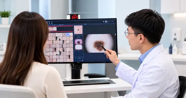
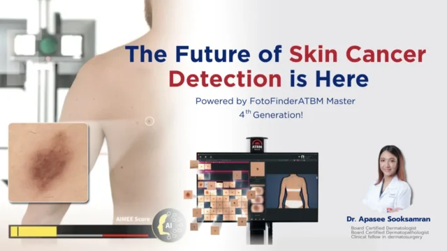
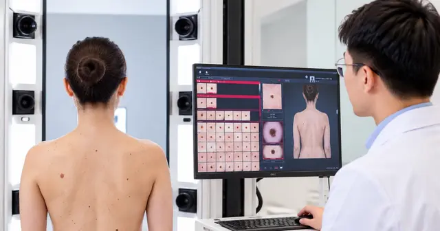
For selected patients, digital imaging systems such as FotoFinder can add another useful piece of information: a photographic record that allows moles and other pigmented lesions to be documented and compared over time.
However, FotoFinder does not replace dermatologist examination or biopsy. If a lesion is sufficiently suspicious for skin cancer, tissue examination may be required to establish the diagnosis.
What Is a Mole Check?
A mole check is a medical assessment of one or more moles or pigmented skin lesions.
Some patients come because they have discovered one particular mole that has changed. Others may have numerous moles and want a broader skin examination.
During the consultation, the dermatologist may assess:
- whether a lesion is new;
- whether it has changed;
- its size, shape and border;
- the distribution of pigment;
- whether it looks different from the patient’s other moles;
- symptoms such as bleeding, crusting, itching or tenderness;
- personal history of melanoma or other skin cancers; and
- relevant family history.
The goal is not to remove every mole.
Instead, the dermatologist determines which lesions appear benign, which may benefit from monitoring, and which require further investigation.
Which Mole Changes Should Be Checked?
A mole should be evaluated if you notice a significant change or something that appears unusual compared with the rest of your skin.
The ABCDE rule provides a useful guide.
A — Asymmetry
One half of the lesion looks different from the other half.
B — Border
The border becomes irregular, poorly defined, notched or uneven.
C — Color
The lesion develops uneven pigmentation or several different colors.
D — Diameter
Larger lesions can be concerning, although size alone cannot determine whether a mole is benign or malignant.
Importantly, melanoma can also be smaller than 6 mm.
E — Evolving
The mole changes in size, shape, color, elevation or other characteristics over time.
The American Cancer Society identifies a new spot or a spot changing in size, shape or color as an important warning sign of melanoma.
Look for the “Ugly Duckling”
The ABCDE rule is useful, but not every melanoma follows it perfectly.
Another practical principle is the ugly duckling sign.
Most people’s moles tend to share a similar appearance. A lesion that looks clearly different from the patient’s other moles may therefore deserve closer examination.
For example, someone may have many small, light-brown moles but develop one dark, irregular lesion.
That difference can be clinically important even if the lesion is relatively small.
Other Suspicious Skin Lesions That Need Examination
A skin cancer check is not limited to dark moles.
Other changes that should be assessed include:
- a sore that does not heal;
- a lesion that repeatedly bleeds;
- persistent crusting;
- a new growing bump;
- a persistent scaly or rough area;
- a changing pink or red lesion;
- an unusual dark area on the palm or sole; and
- a changing pigmented area underneath a nail.
This distinction matters because not every skin cancer looks like a mole.
Basal cell carcinoma and squamous cell carcinoma, for example, can have very different appearances from melanoma.
How Does a Dermatologist Examine a Mole?
The first step is clinical examination.
The dermatologist looks at the lesion and considers it in the context of the patient’s other moles, age, history and risk factors.
When appropriate, the next step may be dermoscopy.
What Is Dermoscopy?
Dermoscopy uses a specialized handheld magnifying instrument called a dermatoscope.
It allows the dermatologist to examine structures and pigment patterns that are difficult to appreciate with the naked eye alone.
Dermoscopy does not involve cutting the skin and is generally painless.
For some lesions, the clinical and dermoscopic appearance may be reassuring.
Others may require photographic monitoring.
And some may be sufficiently suspicious that biopsy is recommended.
Where Does FotoFinder Fit Into a Mole Check?
FotoFinder is a digital imaging system that can be used to document skin lesions and, depending on the system and clinical indication, support photographic comparison over time.
This is particularly useful because a dermatologist is sometimes trying to answer two different questions:
What does this mole look like today?
and
Has this mole changed since the previous examination?
Clinical examination and dermoscopy primarily help answer the first question.
A standardized photographic record can help answer the second.
How FotoFinder Helps Monitor Moles Over Time
Imagine a patient with dozens—or even hundreds—of pigmented lesions.
At a single appointment, the dermatologist can examine suspicious lesions. But remembering the precise size, shape and pigmentation of every mole months or years later is difficult.
Digital documentation creates a baseline.
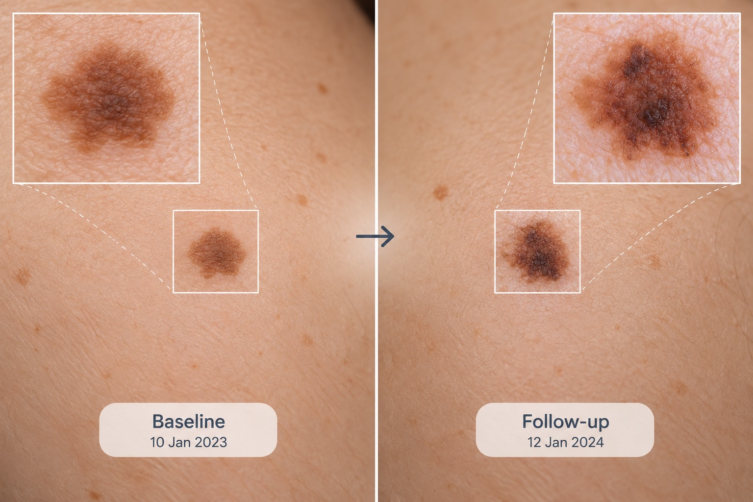
During subsequent examinations, current images can be compared with previous images to look for:
New lesions
A spot may appear that was not present on previous photographs.
Changes in existing lesions
A previously documented mole may have changed in size, shape or pigmentation.
Stable lesions
A lesion may appear similar to its previous documented appearance.
This information is then interpreted by the dermatologist together with the clinical and dermoscopic findings.
Who May Benefit from FotoFinder Mole Monitoring?
Not every patient coming for a mole check requires digital mole mapping.
It may be particularly useful in selected patients with:
- numerous moles;
- multiple atypical moles;
- difficulty keeping track of individual lesions;
- previous melanoma;
- relevant family history of melanoma;
- lesions that the dermatologist considers appropriate for monitoring; or
- other clinical factors that make longitudinal comparison useful.
For someone with one obviously concerning lesion, however, taking photographs and waiting may not be the appropriate approach.
The dermatologist first needs to decide whether the lesion is safe to monitor.
FotoFinder vs Dermoscopy: What Is the Difference?
Patients sometimes assume they are the same thing, but they serve different purposes.
Dermoscopy allows the dermatologist to examine the microscopic-looking structures and pigment patterns within an individual skin lesion.
Digital mole mapping or photographic monitoring creates a record that can be compared over time.
They can therefore complement each other.
For selected patients, the pathway may involve:
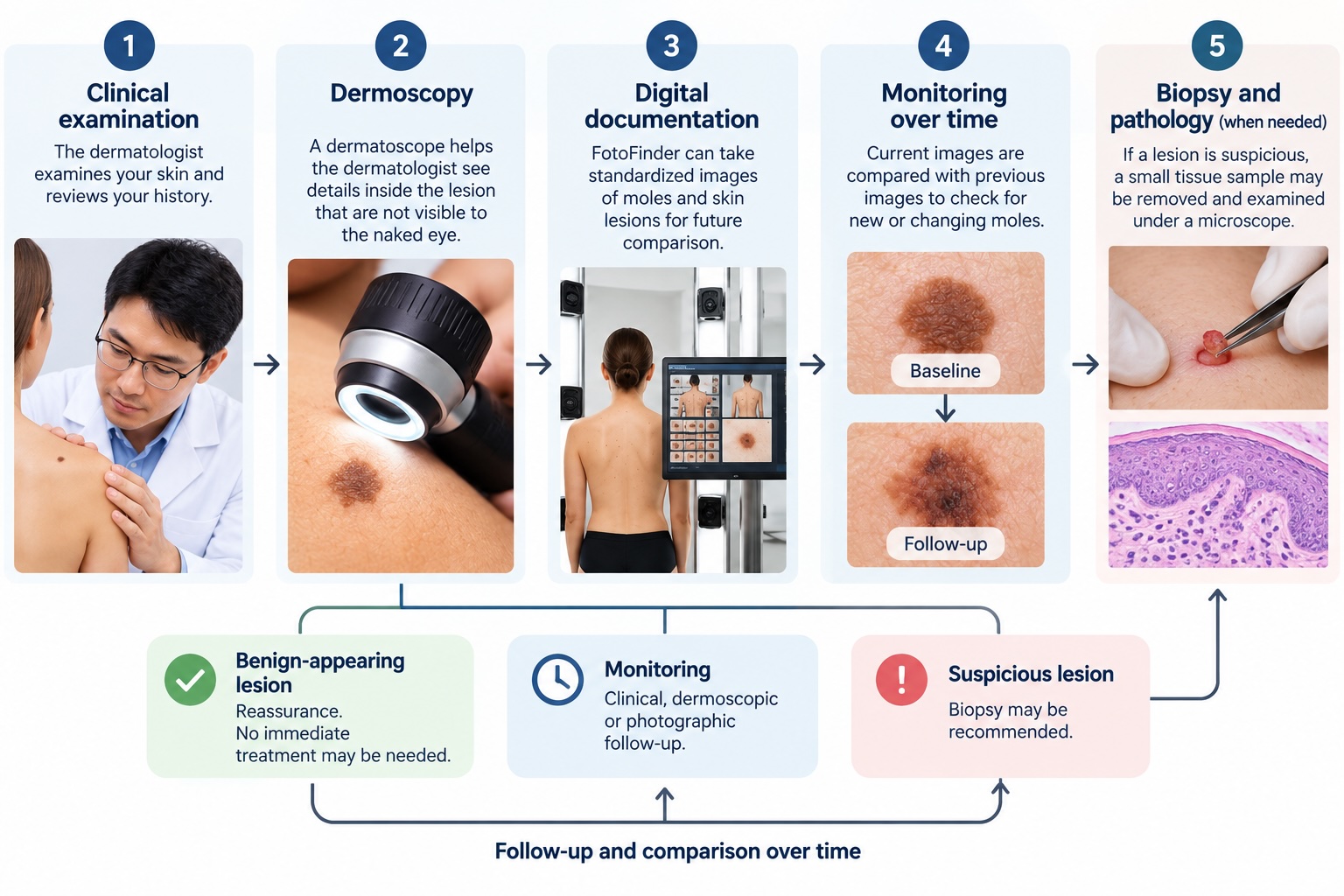
Clinical examination → Dermoscopy → Digital documentation → Comparison at follow-up
But if the lesion is suspicious:
Clinical examination → Dermoscopy → Biopsy when indicated → Pathology
Digital monitoring should not be used to delay appropriate biopsy.
Can FotoFinder Tell Whether a Mole Is Cancerous?
Not by itself.
Digital imaging can help document lesions and identify changes that may warrant closer evaluation, but a photograph or computer-assisted assessment does not provide a definitive tissue diagnosis of melanoma.
The dermatologist interprets the imaging findings in combination with the clinical examination.
If melanoma or another skin cancer is suspected, a biopsy or excision may be required so that the tissue can be examined by a pathologist.
This distinction is important:
FotoFinder helps document and monitor.
The dermatologist assesses.
Pathology confirms the diagnosis when tissue examination is required.
Does a Changing Mole Always Mean Melanoma?
No.
Many benign skin lesions can change gradually, and detecting a difference between two photographs does not automatically mean that the lesion is cancerous.
Likewise, a lesion does not become “safe” simply because a computer does not flag it.
The dermatologist considers multiple factors, including:
- clinical appearance;
- dermoscopic pattern;
- rate and type of change;
- patient’s age;
- location of the lesion;
- other moles on the patient’s body;
- previous skin cancer history; and
- relevant family history.
Digital imaging provides additional information, rather than replacing clinical judgment.
When Is a Biopsy Needed?
A biopsy may be recommended when a lesion cannot be confidently diagnosed by examination alone or when there are features concerning for skin cancer.
During a skin biopsy, tissue is removed and sent for pathological examination under a microscope.
This can determine whether the lesion is benign, melanoma, another type of skin cancer, or another skin condition.
Importantly, being advised to have a biopsy does not mean that the patient definitely has cancer.
It means that tissue examination is needed to establish or clarify the diagnosis.
Monitoring or Biopsy: How Does the Dermatologist Decide?
This is one of the most important decisions during a mole check.
A lesion may broadly fall into one of several pathways:
Benign-appearing lesion
No immediate treatment may be necessary.
Lesion suitable for monitoring
Clinical, photographic or dermoscopic follow-up may be recommended.
Uncertain or suspicious lesion
Biopsy may be appropriate.
Confirmed skin cancer
Treatment is planned according to the diagnosis, location, size and other relevant clinical factors.
The correct pathway depends on the individual lesion.
Why Suspicious Moles Should Not Simply Be Lasered Off
Patients sometimes ask whether an unwanted dark spot or mole can simply be removed with a laser.
For a lesion that has not been confidently diagnosed, this can be problematic.
Destroying a suspicious pigmented lesion may mean that adequate tissue is no longer available for pathological examination.
Therefore, when a mole has unusual or suspicious features:
Diagnosis first. Treatment second.
Once a lesion has been appropriately evaluated and confirmed to be benign, the treatment considerations are different.
Do I Need a Full-Body Mole Check?
Not necessarily.
If you have one new lesion, the dermatologist may focus initially on that specific concern.
A more comprehensive skin examination may be appropriate depending on your history, number of moles and skin cancer risk.
Areas that are difficult to examine yourself can include:
- scalp;
- ears;
- back;
- backs of the legs;
- soles of the feet;
- between the toes; and
- nails.
Melanoma can occur in areas that receive relatively little sun exposure, so patients should not assume that only sun-exposed skin matters.
How Often Should Moles Be Monitored?
There is no universal schedule suitable for everyone.
Follow-up depends on factors such as:
- number and type of moles;
- presence of atypical lesions;
- personal history of melanoma;
- previous skin cancers;
- family history;
- current examination findings; and
- whether a particular lesion needs closer surveillance.
Some patients may only need routine self-monitoring and medical review when something changes.
Others may benefit from scheduled dermatologist examinations and photographic comparison.
The interval should therefore be individualized rather than chosen simply because a certain number of months has passed.
What Should I Do Between Mole Checks?
Become familiar with your skin.
You do not need to diagnose your own moles. Instead, look for meaningful changes.
Pay particular attention to:
- new lesions;
- changing moles;
- lesions that look different from your others;
- non-healing sores;
- repeated bleeding or crusting; and
- unexplained persistent skin growths.
If you notice a concerning change, do not wait until your next scheduled FotoFinder appointment.
Arrange an earlier dermatologist assessment.
Mole Checks for Expats and International Patients in Phuket
Patients living in several countries often have skin examinations performed by different doctors.
Keeping previous records can make comparison easier.
If available, bring:
- previous photographs of the lesion;
- previous digital mole-mapping images;
- pathology reports;
- biopsy results;
- records of previous melanoma or other skin cancers; and
- information about relevant family history.
If you are visiting Phuket temporarily, tell the dermatologist when you plan to leave Thailand.
If biopsy is required, there may need to be enough time for wound care, pathology results, suture removal when applicable, and planning any further treatment.
If you leave before this process is complete, records can be important for continuity of care with your dermatologist at home.
What to Expect During a Mole Check in Phuket
A typical assessment may begin with a discussion about the lesion that concerns you and whether you have noticed any changes.
The dermatologist then examines the lesion and may assess other areas of the skin depending on your history and reason for consultation.
When appropriate, examination may include dermoscopy.
For selected patients, FotoFinder or other standardized digital imaging can be used to establish a baseline and support future comparison.
After the assessment, the dermatologist should explain which pathway is appropriate:
No treatment required
Self-monitoring
Photographic or dermoscopic monitoring
Biopsy and pathology
Treatment or referral if a skin cancer is diagnosed
The objective is not to perform as many tests as possible. It is to choose the appropriate level of investigation for the individual patient and lesion.
When Should You See a Dermatologist Promptly?
Consider arranging an assessment rather than waiting for routine screening if you notice:
- a rapidly changing mole;
- significant change in color or shape;
- an irregular new pigmented lesion;
- repeated spontaneous bleeding;
- persistent ulceration;
- a sore that does not heal;
- a lesion that looks markedly different from your other moles; or
- a suspicious lesion on the palm, sole or underneath a nail.
A photograph can be useful for documenting a lesion, but a concerning change should be examined rather than simply photographed and watched at home.
Frequently Asked Questions
Is FotoFinder better than a regular mole check?
They are not alternatives. A dermatologist examination evaluates the lesion clinically, while digital imaging can provide a record for comparison over time. Some patients benefit from both.
Can FotoFinder diagnose melanoma?
No. FotoFinder can assist with documentation and monitoring, but melanoma diagnosis may require biopsy and pathological examination.
Do I need FotoFinder if I only have one mole?
Not necessarily. A dermatologist may simply examine the lesion and use dermoscopy. Digital monitoring is more useful in selected situations.
Is FotoFinder useful if I have many moles?
It can be particularly useful when there are numerous lesions and longitudinal comparison would help the dermatologist monitor for new or changing lesions.
What happens if FotoFinder shows that a mole has changed?
The dermatologist assesses the type of change together with the clinical and dermoscopic appearance. Depending on the findings, further monitoring or biopsy may be recommended.
Does every changing mole need to be removed?
No. Change does not automatically mean cancer. The significance of the change needs to be interpreted by a dermatologist.
Can a small mole be melanoma?
Yes. Melanoma can occur in lesions smaller than the commonly referenced 6 mm threshold. Size should never be considered in isolation.
Can I have a mole checked while on holiday in Phuket?
Yes. A new or suspicious lesion can be medically evaluated while travelling. If further investigation is needed, your travel schedule should be discussed so that follow-up can be planned appropriately.
Should I remove a suspicious mole with a laser?
A suspicious pigmented lesion should be diagnosed before destructive cosmetic treatment. If melanoma is a possibility, tissue may be needed for pathological examination.
Should I wait for my next mole-mapping appointment if something changes?
No. A new or significant change should be assessed promptly rather than automatically waiting for the next scheduled imaging appointment.