A mole may look harmless when examined today, but sometimes the most useful information is how it changes over time.
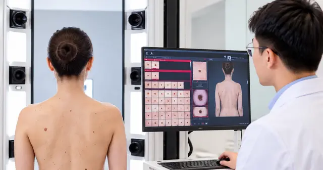
Digital mole mapping uses standardized skin photography and, depending on the system and clinical indication, detailed images of selected moles to create a visual baseline. These images can then be compared at future visits to help a dermatologist identify new lesions or changes in existing moles.
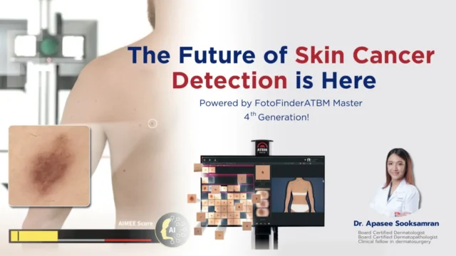
FotoFinder is one system used for this type of photographic skin surveillance. Importantly, digital mole mapping does not replace a dermatologist examination, diagnose melanoma by itself, or replace a skin biopsy when cancer is suspected. Total-body photography is particularly useful as a surveillance tool in selected people at higher risk, including some patients with numerous or atypical moles.DermNet®
For patients living in Phuket—or international patients spending extended periods in Thailand—having a consistent photographic record can also make future mole comparisons more systematic.
What Is Digital Mole Mapping?
Digital mole mapping is a method of documenting the skin with standardized photographs so that the appearance and distribution of moles can be compared over time.
Depending on the system being used, this may include:
- standardized photographs of different areas of the body;
- a baseline record showing the location of existing moles;
- closer photographs of selected lesions;
- dermoscopic images of particular moles when clinically appropriate; and
- comparison with images obtained during subsequent examinations.
FotoFinder systems can support total-body mapping workflows that create and evaluate whole-body images and maintain a timeline for comparison.FotoFinder Systems
The important concept is comparison.
Instead of relying only on memory—or on photographs taken under different lighting, distances and angles—the dermatologist can compare standardized images from different points in time.
Why Is Tracking Moles Over Time Useful?
Many benign moles remain relatively stable for years. A lesion that is new or evolving, however, deserves attention.
Changes that may be important include:
- increasing size;
- changing shape;
- development of an irregular border;
- changing or uneven pigmentation;
- a mole becoming noticeably different from the patient’s other moles; or
- development of symptoms such as bleeding, crusting, itching or pain.
A new lesion or a lesion changing in size, shape or color is an important warning sign for melanoma. Another useful clue is the “ugly duckling” sign—a spot that looks noticeably different from the person’s other moles.American Cancer Society
Digital mole mapping gives the dermatologist an additional piece of information:
What did this lesion look like previously?
That can be particularly useful when a person has many moles and remembering the appearance of every individual lesion is difficult.
Who May Benefit from Digital Mole Mapping?
Digital mole mapping is not necessarily required for everyone.
It may be particularly useful when a dermatologist considers a patient to have an increased need for photographic surveillance, for example someone with:
- numerous moles;
- multiple atypical or dysplastic moles;
- a previous melanoma;
- a strong family history or other recognized melanoma risk factors;
- moles that are difficult to monitor individually;
- lesions that the dermatologist has specifically decided to observe; or
- difficulty performing reliable self-examination.
Photographic skin surveillance, including total-body photography, is particularly considered for higher-risk individuals with high mole counts or dysplastic nevi.DermNet®
The decision should still be individualized. A patient with one concerning lesion may need a focused dermatologist examination—and potentially a biopsy—rather than simply photographing the lesion and waiting.
What Happens During a FotoFinder Mole Mapping Examination?
The exact workflow depends on the FotoFinder system available and the dermatologist’s assessment, but a mole-mapping visit generally involves several steps.
1. Medical and Skin Cancer History
The dermatologist may ask about:
- a previous melanoma or other skin cancer;
- previous abnormal or dysplastic moles;
- family history of melanoma;
- significant previous sun exposure or sunburn;
- whether a particular mole is new or changing;
- symptoms such as bleeding, itching or pain; and
- previous biopsies or pathology results.
Medical history remains an important part of assessing a suspicious skin lesion.American Cancer Society
For international patients, bringing previous photographs or pathology reports can be particularly useful.
2. Dermatologist Skin Examination
Digital imaging should not be viewed as a replacement for the dermatologist.
The doctor examines the skin and determines which lesions appear benign, which may be suitable for monitoring, and which require closer assessment.
Depending on the reason for the appointment and the patient’s risk profile, the examination may focus on one lesion or involve a more extensive skin examination.
3. Standardized Photography
Images are taken to create a baseline record of the skin.
The purpose is not simply to take photographs. Standardization makes later comparisons more meaningful. 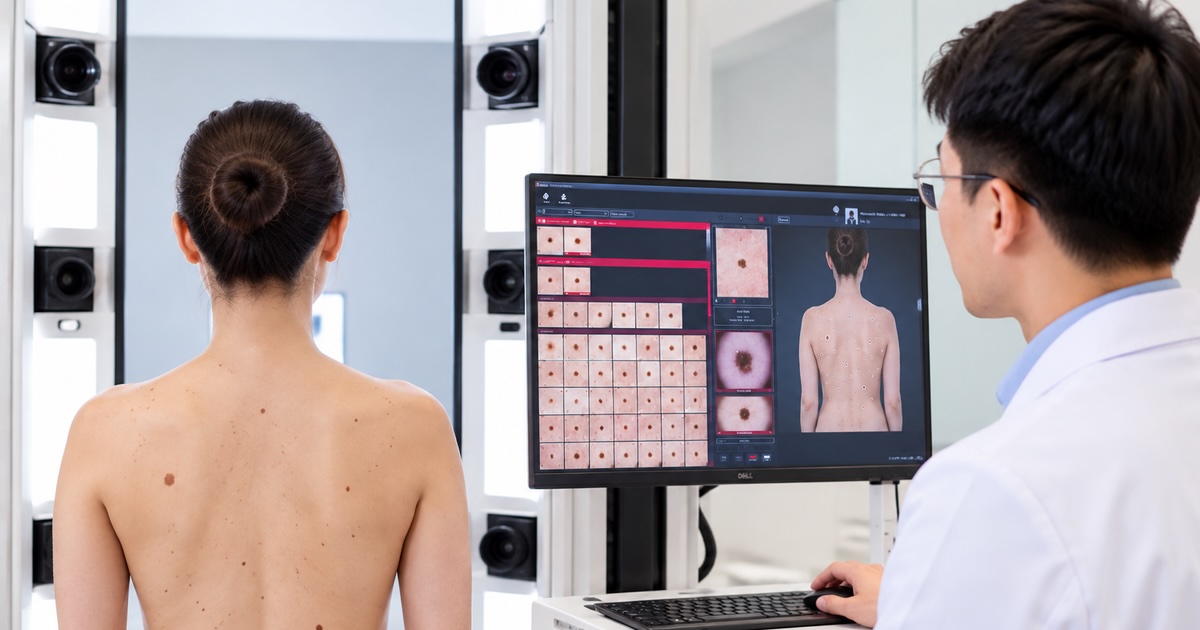
At a future examination, previous and current images can be reviewed to look for new lesions or visible changes.
4. Dermoscopy When Appropriate
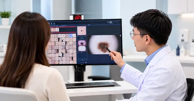
A dermatologist may examine selected moles using dermoscopy.
A dermatoscope is a specialized magnifying device that allows structures and pigment patterns beneath the skin surface to be examined more clearly than with the naked eye.
Digital dermoscopic images of selected lesions can also be stored for comparison when monitoring is considered clinically appropriate.
5. A Management Decision
After examination, a lesion may be considered:
Benign appearing → suitable for routine observation
Suitable for photographic or dermoscopic monitoring → follow-up according to the dermatologist’s assessment
Suspicious → further evaluation or biopsy may be recommended
This distinction matters because not every mole needs to be removed, but not every suspicious mole should simply be monitored.
FotoFinder Mole Mapping vs a Regular Mole Check
They are related, but they are not the same thing.
A regular mole check is primarily a clinical examination. The dermatologist assesses the appearance of the patient’s moles and other skin lesions at that visit, often using dermoscopy when appropriate.
Digital mole mapping adds a longitudinal photographic record.
That makes it particularly useful when the clinical question is:
Has this mole changed since the previous examination?
For some patients, combining clinical examination, dermoscopy and photographic comparison can provide more information than a single examination alone.
However, the technology supports—not replaces—clinical judgment.
Can FotoFinder Detect Melanoma?
This question needs careful wording.
FotoFinder images can help a dermatologist identify and monitor changes that may warrant further assessment, but imaging alone does not establish a definitive diagnosis of melanoma.
A suspicious lesion may need to be removed or sampled and examined under a microscope by a pathologist.
DermNet specifically notes that while photographic surveillance can help monitor new or changing lesions, a lesion showing clinical or dermoscopic evidence concerning for melanoma should be excised rather than simply continuing observation.DermNet®
In other words:
Digital imaging helps identify change.
Dermatologists interpret the findings.
Pathology establishes the tissue diagnosis when biopsy is required.
When Should a Mole Be Biopsied Instead of Monitored?
This is one of the most important limitations of mole mapping.
Monitoring is appropriate only when the dermatologist believes observation is reasonable.
If a lesion is sufficiently suspicious for melanoma or another skin cancer, the safer next step may be a biopsy rather than waiting for another set of photographs.
A biopsy allows tissue to be examined microscopically and can determine whether the lesion represents melanoma, another skin cancer, or a benign condition.American Cancer Society
Digital mole mapping should therefore never be used to delay biopsy of a clinically suspicious lesion.
What Mole Changes Should You Watch For?
The ABCDE rule is a useful way to remember some melanoma warning signs.
A — Asymmetry
One half looks different from the other.
B — Border
The edges become irregular, blurred, ragged or poorly defined.
C — Color
There are different colors or an uneven distribution of pigmentation.
D — Diameter
Larger lesions can be concerning, although this rule has an important limitation: melanoma can be smaller than 6 mm.
E — Evolving
The mole changes in size, shape, color or other characteristics.
“Evolving” is particularly relevant to digital mole mapping because comparison over time can help document change.American Cancer Society
The “Ugly Duckling” Sign
Not every melanoma follows the classic ABCDE pattern.
Another useful principle is to look for a lesion that appears different from the patient’s other moles.
For example, a person may have dozens of similar light-brown moles but develop one very dark, irregular lesion.
That outlier deserves attention even if it is relatively small. The American Cancer Society identifies a spot that looks different from the person’s other spots as another important melanoma warning sign.American Cancer Society
Does Every Changing Mole Mean Melanoma?
No.
Moles and other skin lesions can change for benign reasons, and a change seen between photographs does not automatically mean cancer.
Likewise, not every mole that looks unusual is melanoma.
The purpose of monitoring is to give the dermatologist additional information that can be interpreted alongside:
- the clinical appearance;
- dermoscopic features;
- the patient’s age and history;
- the location of the lesion;
- the pattern of the patient’s other moles; and
- relevant skin cancer risk factors.
The decision to continue monitoring or perform a biopsy should be made clinically rather than by photography alone.
What Are the Limitations of Digital Mole Mapping?
Digital mole mapping is useful, but patients should understand what it cannot do.
It cannot diagnose every skin cancer
Not all skin cancers are pigmented moles. Basal cell carcinoma and squamous cell carcinoma may appear as non-healing sores, scaly areas, crusted lesions or new growths.
It does not replace pathology
If tissue diagnosis is necessary, a biopsy is still required.
It does not replace examination by a dermatologist
Images need clinical interpretation.
Not every lesion is appropriate for monitoring
A suspicious or rapidly changing lesion may require immediate investigation rather than waiting for a future comparison.
It cannot guarantee that melanoma will be detected
No screening or imaging method can provide a guarantee.
These limitations are important when patients see terms such as “AI mole analysis.” Automated tools may assist with assessment, but they should not be interpreted as a standalone cancer diagnosis. The American Cancer Society similarly notes ongoing concerns regarding the accuracy of consumer AI skin-cancer tools and recommends professional evaluation of concerning lesions.American Cancer Society
How Often Should Digital Mole Mapping Be Repeated?
There is no single interval that is appropriate for everyone.
The appropriate follow-up schedule depends on factors such as:
- number and type of moles;
- previous melanoma or other skin cancer;
- family history;
- atypical mole history;
- findings during the current examination; and
- whether a particular lesion requires short-term surveillance.
Melanoma follow-up intensity should be individualized according to factors including disease stage and risk of recurrence or a new primary melanoma.American Academy of Dermatology
Your dermatologist should recommend the appropriate interval based on your individual findings rather than following an arbitrary schedule.
Do I Still Need to Check My Own Skin?
Yes.
Digital mole mapping is performed at specific points in time. Changes can develop between appointments.
Become familiar with your normal pattern of moles and skin lesions and look for anything new, changing or noticeably different.American Cancer Society
Do not wait for your next scheduled mole-mapping appointment if you notice a concerning change.
When Should You See a Dermatologist Promptly?
Arrange an assessment if you notice:
- a new mole or unusual pigmented lesion;
- a mole changing in size, shape or color;
- a lesion that looks very different from your other moles;
- recurrent bleeding or crusting;
- a sore that does not heal;
- a new lump or growth;
- new persistent itching, tenderness or pain in a lesion; or
- a changing dark area on the palm, sole or underneath a nail.
Melanoma can occur in areas that receive little sun exposure, including the palms, soles and beneath nails.American Cancer Society
If a lesion is changing rapidly or already appears suspicious, do not wait for the next routine photographic follow-up.
Mole Screening for Expats and International Patients in Phuket
Digital records can be particularly useful for people who live between different countries.
If you have previously undergone mole mapping, skin biopsy or treatment for skin cancer outside Thailand, consider bringing:
- previous mole photographs;
- digital mole-mapping records if available;
- pathology reports;
- previous biopsy or surgery reports;
- history of melanoma or other skin cancers; and
- information about relevant family history.
If you are visiting Phuket temporarily, tell the dermatologist your departure date.
If a lesion needs biopsy, follow-up may involve wound care, pathology results, possible suture removal and, depending on the diagnosis, additional treatment. Planning these steps before travelling can make continuity of care easier.
Should a Suspicious Mole Be Removed with a Laser?
A pigmented lesion should be properly diagnosed before cosmetic destruction if its nature is uncertain.
Laser treatment destroys tissue and may leave insufficient material for pathological examination.
If a mole or pigmented lesion has suspicious features, the priority is:
Diagnosis first → appropriate treatment second.
This is different from treating a lesion that has already been confidently assessed as benign.
Digital Mole Mapping and Skin Cancer Check in Phuket
For patients seeking a skin cancer check or mole screening in Phuket, the appropriate assessment depends on the individual.
Some patients simply need a dermatologist to examine one new or changing lesion.
Others may benefit from a more comprehensive skin examination.
Patients with numerous or atypical moles may, in selected circumstances, benefit from digital mole mapping to establish a baseline for future comparison.
And when a lesion is suspicious, the appropriate next step may be biopsy and pathological examination rather than continued photographic monitoring.
The technology is therefore most useful when it forms part of a broader clinical pathway:
Dermatologist examination → dermoscopy when appropriate → digital monitoring when appropriate → biopsy when indicated → pathology → treatment or follow-up according to the diagnosis.
Frequently Asked Questions
Is FotoFinder the same as a skin cancer test?
No. FotoFinder can provide standardized images that help document and compare skin lesions over time. It does not replace dermatologist assessment or biopsy when tissue diagnosis is required.
Can FotoFinder tell whether a mole is melanoma?
It can help document features and changes that a dermatologist may consider suspicious, but a definitive melanoma diagnosis generally requires pathological examination of tissue obtained by biopsy or excision.American Cancer Society
Is digital mole mapping useful if I have many moles?
Potentially. Total-body photography is particularly useful in selected higher-risk people, including those with a high number of moles or dysplastic nevi.DermNet®
Does every mole need to be photographed?
No. The appropriate imaging strategy depends on the patient’s skin, risk factors and clinical findings.
What happens if one of my moles changes?
A dermatologist should assess the change. Depending on its clinical and dermoscopic appearance, the lesion might be monitored further or biopsy/excision may be recommended.
Can a small mole be melanoma?
Yes. Although diameter is included in the ABCDE rule, melanomas can be smaller than 6 mm.American Cancer Society
Should I wait until my next FotoFinder appointment if a mole changes?
No. A new or changing lesion should be assessed rather than automatically waiting for the next scheduled imaging session.
Can tourists have their moles checked while in Phuket?
Yes, a concerning lesion can be evaluated while travelling. If biopsy or further treatment is required, the patient’s travel schedule should be considered when arranging pathology, wound care and follow-up.
Can digital mole mapping replace a full skin examination?
No. It is an additional surveillance tool. Clinical examination by a dermatologist remains important.
Should I bring previous mole photographs from another clinic or country?
Yes. Previous images, pathology reports and skin cancer records can provide useful historical information, especially if a particular lesion has changed.