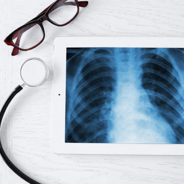
Lung disease is a common malady. It can be identified from the symptoms, blood test, sputum culture and chest x-ray. In most cases, the x-ray is enough for the doctor to diagnose the disease and provides a treatment. Sometimes the doctor may need more detail and will order a computer scan (CT scan) or a pulmonary function testing. To further clarify the diagnosis, a biopsy may be required through bronchoscopy or, if the target sample is inside the lung, thoracotomy.
Lately, with advancements in medical technology, needle aspiration – or biopsy – has become increasingly more common in retrieving a tissue sample for testing. If the lesion is small, the doctor may utilize ultrasound or CT scan to guide the needle into the lesion for more effective result. However, if needle aspiration is not possible, the surgeon will have to perform a thoracoscopy in which a thoracoscope is inserted through a small incision in the chest area to extract a tissue sample from the lesion. The handling of such a case is similar to treating lung diseases where medication is generally prescribed and surgery is less common. Usually, the surgery is needed in the case of a tumor, especially lung cancer, and traditional method would require an open chest operation to remove the growth. Other cases include repairing pneumothorax, removing or treating pulmonary blebs and bullae.
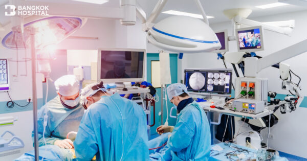
What is Thoracotomy?
Thoracotomy is a surgical procedure to gain access into the thoracic organs that includes heart, lungs and others. There are generally 2 methods for thoracotomy: median sternotomy and lateral approach.
Lung Resection: First of all, it is necessary to understand the anatomy of the lung. The trachea – main air passage way – branches into the left and right lungs. The split pathways feed into various lobar bronchi as each lung is divided into lobes – 3 in the right lung and 2 in the left. Then the air passages further divide into smaller branches to feed the segments within each lobe of which there are 10. For the left lung, there are 5 segments in each of the upper and lower lobes. For the right lung, the upper lobe has 3 segments, the middle lobe has 2, while the lower lobe has 5.
For thoracic surgery, the surgeon will determine how much tissue has to be removed. This depends on the size and characteristic of the lesion, as well as its extent within the lung. Another consideration is its proximity to a bronchus. If the size is small and there is no connection to a bronchus, then wedge resection or biopsy is possible. However, if the lesion is at a segment of a lobe, a segmental resection will be required. Similarly, if it is on the airway to a lobe, then lobectomy will be performed. Otherwise, if the lesion is on the main pathway to a lung, pneumonectomy – where an entire lung is removed – will be needed.
Video Assisted Thoracic Surgery (VATS)
Presently, video assisted thoracic surgery has become more widespread and is performed instead of the traditional thoracotomy, especially for segmental resection. However, for an experienced surgeon, it can be performed in lobectomy also. Compared to thoracotomy, VATS yields smaller incisions and reduced risks, as well as less pain and shorter hospitalization with less complication after the surgery – no more than 5% as compared to as much as 30% – and the patient can return to work sooner. In addition, VATS technique has been utilized in other types of procedures that include gastrointestinal, spinal, and cardiac surgeries.