Kidney stones, also known as renal calculi or nephrolithiasis are hard deposits made of minerals that form inside the kidneys. Although kidney stones can strike anyone at any age, men are more susceptible to develop this condition than women. Peak incidence occurs between ages 30 to 40 years. If they are left untreated, serious complications might potentially develop, including infection of the kidney, sepsis, injury to ureter and impaired kidney function that eventually leads to chronic kidney disease. Knowing the warning signs of kidney stone allows for an early diagnosis and effective treatment given in a timely manner before the condition severely progresses.
Get to know kidney stones
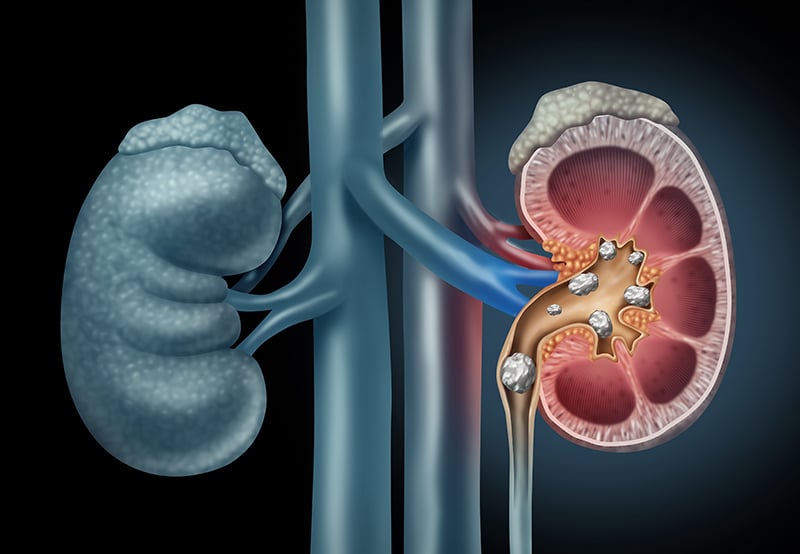
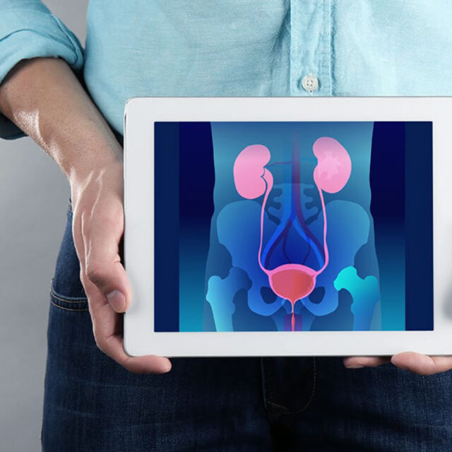
Kidney stones are defined as hard deposits made of minerals that form inside the kidneys. These stones often form when the urine becomes concentrated, allowing minerals to crystallize and stick together. Kidney stones can affect any part of the urinary tract, extending from the kidneys to bladder. Kidney stones usually range in size from as small as a grain of sand to the size of a golf ball. More importantly, kidney stones can recur after receiving treatments.
Risk factors
Factors related to high amount of calcium in the urine increase the risk of developing kidney stones, these include:
- Consuming a diet that is high in calcium, protein, sodium (salt) and sugar. An excessive salt in the diet increases the amount of calcium that the kidneys must filter.
- Not drinking enough water.
- Adding more sugar into the beverages.
- Eating foods e.g. nuts, bamboo shoot, chocolate, spinach and sweet potato that contain high amount of oxalate, a substance that prohibit calcium absorption.
- Taking high dose of vitamin C, greater than 1,000 mg. per day.
- Overactive parathyroid glands affecting calcium levels.
- Complication of gout, resulting in uric acid buildup in the blood and crystal formation in the joints and the kidneys.
- Digestive diseases e.g. inflammatory bowel disease or chronic diarrhea that cause changes in the digestive process affecting calcium and water absorption.
- Being overweight or obesity.
Warning signs and symptoms
A kidney stone usually does not cause symptoms until it moves around within the kidney or passes into the ureters. If it becomes lodged in the ureters, it may block the flow of urine causing the kidney to swell and the ureter to spasm. Signs that might likely indicate kidney stones are as follows:
- Severe and sharp pain in the side and back, below the ribs
- Pain that radiates to one side of the back or lower abdomen
- Pain that comes in waves with fluctuating intensity
- Fever and chills, if an infection is present
- Nausea and vomiting
- Cloudy, pink, red or brown urine
- Urination present with sand-like particles
- Pain or burning sensation while urinating
- Urinating more often than usual
- Urinating in small amounts
- Inability to urinate
- Intense pain in the abdomen (renal colic) when the stone moves to the ureters
**Nevertheless, some patients may not develop any of these symptoms.
Diagnosis
If kidney stones are suspected, additional tests including imaging tests must be conducted by expert specialists in order to make a confirmative diagnosis. Diagnostic tests and procedures include:
- Urine examination. The presence of red blood cells in the urine might be presumably associated with kidney stones.
- Blood testing. Blood tests are used to reveal calcium and uric acid levels in the blood as well as other relevant parameters. Patients diagnosed with kidney stones usually have an excess of blood calcium and uric acid levels.
- Abdominal X-rays. X-rays can be deployed to detect kidney stones formed in the urinary tract.
- Computerized tomography (CT) scan. CT scan helps reveal tiny stones.
- Kidney ultrasound. Kidney ultrasound is a noninvasive test that aims to detect stones in the kidney.
- X-ray with IVP (intravenous pyelogram). IVP is an x-ray exam that uses intravenous contrast material to evaluate the kidneys, ureters and bladder and help determine the cause of kidney stones, resulting in an appropriate treatment plan and prevention of kidney stone recurrence.
*** Additional tests or procedures might be further required in some cases, based upon specialist’s recommendations.
Treatment
Treatment for kidney stones varies, depending on the type of stone and the cause. Treatment options include:
- Non-surgical treatments: Small stones with mild symptoms usually does not require invasive treatment. To pass small stones through urination, it is highly recommended to drink a lot of water. In some cases, the the specialist might consider proscribing certain medications that relax the muscles in the ureter, helping patients to pass the stones more quickly and with less pain.
- Using sound waves to break up stones (extracorporeal shock wave lithotripsy or ESWL): ESWL uses sound waves to create strong vibrations, known as shock waves, that break the stones into tiny pieces, allow them to be passed through urination. It is advised to treat kidney stones sized smaller than 2 cm. Since the procedure can cause mild to moderate pain and some complications, therefore it should be performed by highly experienced urological surgeons.
- Using a scope to remove stones (ureteroscope): To remove a small stone sized smaller than 3 cm, the urologist might pass a thin lighted tube (ureteroscope) equipped with a camera through the urethra and bladder to the ureter. Once the stone is located, special tools can snare the stone, breaking it into pieces that will pass in the urine.
- Surgery to remove large stones in the kidney (percutaneous nephrolithotomy): To surgically remove a large kidney stone that failed to other treatments, percutaneous nephrolithotomy might be considered. This procedure involves surgically removing a large kidney stone using small telescopes and surgical instruments inserted through a small incision made in the back. Posing some procedure related risks, this surgical treatment must be only conducted by highly experienced and well trained urological surgeons.
Prevention
- Drink sufficient water throughout the day to prevent the crystallization of minerals and salt in the urine.
- Continue eating calcium-rich foods, but use caution with calcium supplements.
- Minimize the consumption of animal protein and daily products e.g. milk and butter.
- Eat more vegetable and fewer oxalate-rich foods.


