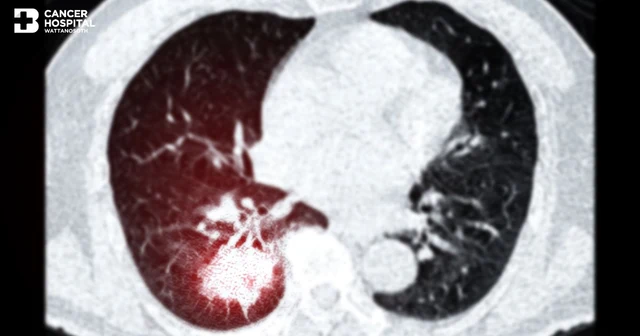

Lung cancer (Lung Cancer) occurs when epithelial cells lining the inside of the lungs divide abnormally and rapidly until they become a malignant tumor that can invade nearby tissues or spread to other organs. It is one of the leading causes of cancer-related death among Thai people and worldwide. A major problem is that most patients are unaware and have no symptoms until the cancer has already progressed or metastasized. Screening with Low-Dose Chest CT Scan therefore helps detect small spots in the lungs that may be early-stage lung cancer even before symptoms appear, reducing lung cancer mortality, increasing the chance of cure, and also reducing lung cancer recurrence.
How the lungs work
The lungs are the main organs of the respiratory system. The smallest components of the lungs are hundreds of millions of tiny air sacs (Alveoli) that are delicate and sensitive to environmental irritants, and they function to exchange oxygen and carbon dioxide. The tissue of the alveoli has no sensory receptors that would cause early abnormal symptoms, so lung cancer is often discovered at stages 3–4, when the tumor has spread to larger airways, invaded nearby organs, or already metastasized to other organs. This makes treatment more difficult, lowers the chance of cure, and increases cancer mortality. Proactive screening with Low-Dose Chest CT Scan is therefore important, especially for those in high-risk groups for lung cancer; however, individuals without risk factors can also undergo proactive screening.
What is Low-Dose Chest CT Scan?
Low-Dose Chest CT Scan is a low-radiation computed tomography (CT) scan in which image quality remains sufficient to diagnose lung masses that are less than or equal to 2 millimeters. The person being examined receives less radiation than with a conventional chest CT scan (Chest CT Scan) by 70–80%. This technology is endorsed by leading global oncology organizations such as the United States Preventive Services Task Force (USPSTF), American Cancer Society (ACS), and the National Comprehensive Cancer Network (NCCN), European Guidelines, and others, as a standard screening test for lung cancer in high-risk groups.
Differences between Low-Dose CT Scan and other tests
|
Test modality |
Detection capability |
Radiation dose |
Features |
|
Low Dose CT (LDCT) |
Lung nodules or masses of any size, regardless of appearance (ground-glass / part-solid–solid/solid), can be seen. |
~1–1.5 mSv |
|
|
Standard Chest CT |
~6–7 mSv |
|
|
|
Chest X-Ray |
Masses that are solid-appearing or large |
~0.1 mSv |
|
|
PET-CT Scan |
Tissues with high glucose uptake Usually a whole-body examination |
~25 mSv |
|
|
Photon-Counting Detector CT |
Can also be used to evaluate abnormalities in the chest |
~1.5 – 4 mSv |
|
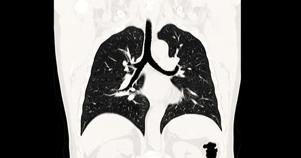
What can Low-Dose Chest CT Scan detect
- Masses or spots in the lungs or chest cavity, both benign and malignant
- Pneumonia/infection
- Emphysema
- Pleural effusion from various causes such as inflammation, infection, cancer
- Thoracic aortic aneurysm
- Coronary artery calcification
Warning signs of lung cancer
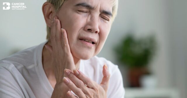
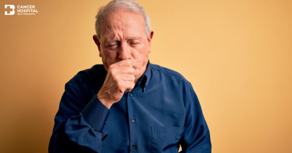
Lung cancer often has no symptoms in the early stage, so screening with Low-Dose Chest CT Scan is important. However, as lung cancer progresses, the following symptoms may occur:
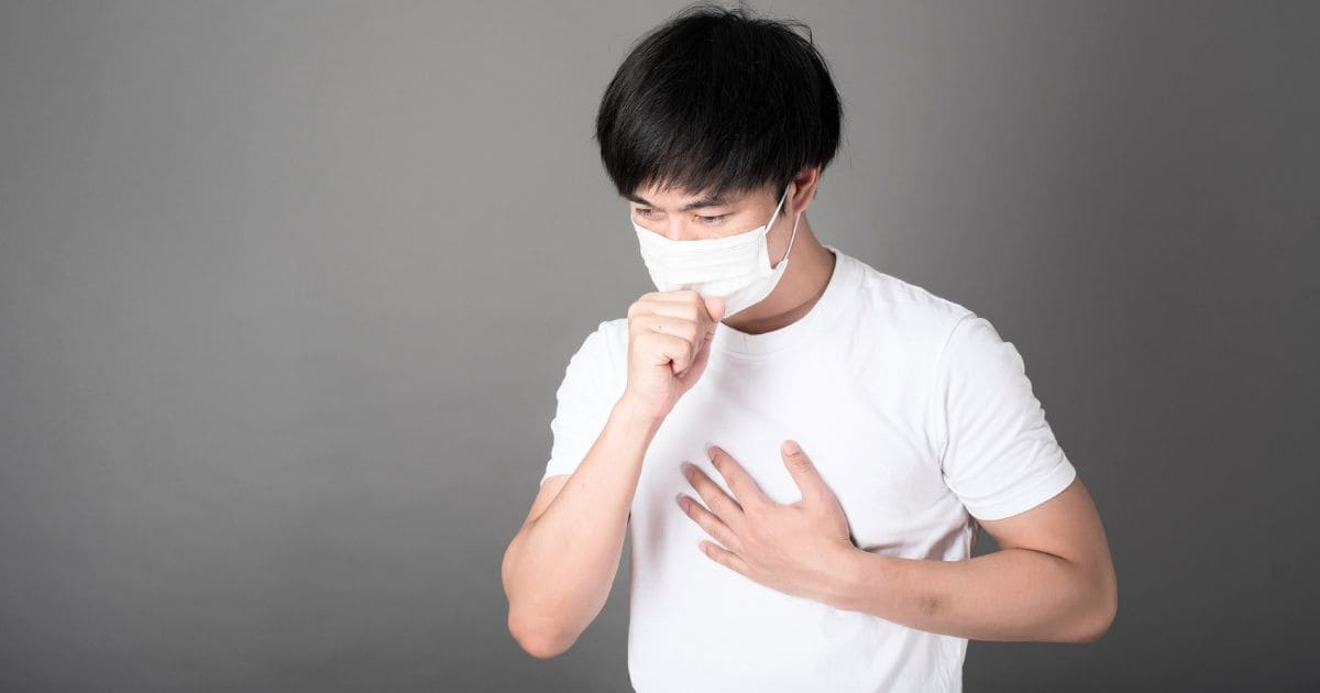
- Symptoms from a tumor growing in the lungs (often stage 3) include chronic cough, coughing up blood, hoarseness, persistent voice change
- Symptoms when cancer has spread to other organs (often stage 4) include shortness of breath, difficulty breathing, bone pain, fatigue, easy exhaustion, unexplained weight loss
Who should undergo Low-Dose Chest CT Scan
In Asia and other Southeast Asian countries, studies have found that lung cancer can be detected even in non-smokers due to EGFR gene mutations, as well as possibly greater exposure to risk factors that cause lung cancer than in the Americas or Europe, such as toxic particulate matter (PM 2.5), incense smoke, smoke from household combustion, etc. Therefore, annual lung cancer screening with low-dose chest CT is important.
- Men aged 50 years and above, or women aged 45 years and above
- Those with a family history of lung cancer
- Current or former smokers with at least 20 Pack-years
*Pack-years is a unit measuring “cumulative smoking exposure”, calculated as the number of packs smoked per day × the number of years smoked. For example, 1 pack/day for 20 years = 1 × 20 = 20 pack-years or 2 packs/day for 10 years = 2 × 10 = 20 pack-years, etc.
Moderate-risk group who should undergo Low-Dose Chest CT Scan
- Those who live or work in areas with high air pollution, such as PM2.5 dust
- Those who have previously received radiotherapy to the chest
- Those with a history of chronic lung disease
- Those who have previously had other types of cancer
In addition, the general public, even without risk factors, can also undergo screening with Low-Dose Chest CT Scan.
Preparation before Low-Dose Chest CT Scan
- Eat as usual; no need to fast from food or water
- Remove metal jewelry such as necklaces and earrings
- Wear comfortable clothing
- If you are pregnant, inform the doctor or relevant staff

Steps for Low-Dose Chest CT Scan at Bangkok Hospital
- Pre-exam assessment: The doctor takes a risk history and evaluates suitability for the examination.
- Scanning: Lie flat on the CT machine; it takes no more than 10–15 minutes; no contrast injection is required.
- Image analysis: By a radiologist specialized in interpreting chest imaging
- Reporting of results
- A lung cancer specialist explains the results in detail and creates an individualized care and treatment plan.
Treatment options when an abnormality is found
If an abnormality is detected, the multidisciplinary medical team (Multidisciplinary Team: MDT) will jointly plan an individualized treatment approach as follows:
- Tissue diagnosis (Tissue Diagnosis) can be performed in several ways depending on patient suitability and the location of the detected abnormality, such as bronchoscopy (Bronchoscopy), percutaneous biopsy (Trans-Thoracic Needle Biopsy), or minimally invasive thoracoscopic surgery (Minimally Invasive Thoracoscopic Surgery).
- Treatment planning by the multidisciplinary team (Multidisciplinary Team: MDT) to select the most appropriate method and maximize patient benefit. There are multiple options:
- Video-assisted thoracoscopic surgery Video-Assisted Thoracoscopic Surgery (VATS) or robotic-assisted thoracoscopic surgery Robotic-assisted Thoracoscopic Surgery (RATS) primarily for stage 1 lung cancer, and also stages 2–3 in which surgery is combined with other treatments. Thoracoscopic surgery is considered a mainstay treatment with good outcomes, faster recovery, shorter hospital stay, smaller incisions, less pain, and a cure rate as high as 80–90% in early-stage cancer.
- Stereotactic Body Radiation Therapy (SBRT) is used to treat stage 1 lung cancer when patients do not undergo surgery or are not suitable surgical candidates, to treat stage 3 lung cancer, as postoperative treatment in some cases, and for symptom control in advanced cancer. With SBRT or SABR (Stereotactic Ablative Radiotherapy), high-energy radiation is delivered precisely to the tumor in a small number of sessions (1–8 sessions), reducing side effects to surrounding normal tissue.
- Systemic therapy (Systemic Therapy) includes chemotherapy (Chemotherapy), immunotherapy (Immunotherapy), and targeted therapy (Targeted Therapy). Current lung cancer drug treatment includes adjuvant therapy after surgery in certain stage 1 cancers, neoadjuvant or adjuvant therapy in stage 2 cancers to increase the chance of cure, concurrent therapy with radiation in stage 3 cancers, and medical treatment to control stage 4 cancer. Before systemic therapy, gene testing is often performed to analyze genetic alterations such as EGFR, ALK, ROS1, KRAS G12C, PD-L1, and others, helping doctors select drugs appropriately tailored to each patient’s cancer.
Lung cancer prevention and care
- Stop smoking or avoid being near smokers, which significantly reduces the risk of lung cancer
- Avoid areas with high air pollution; if unavoidable, protect yourself by wearing a standard mask or an N-95 mask
- During periods of high air pollution, use an indoor air purifier in a well-sealed room with a HEPA (High-Efficiency Particulate Air) filter. High-efficiency air filters can trap particles as small as 0.3 microns at 99.97% efficiency, such as PM2.5 dust, pollen, dust mites, mold spores, bacteria, and some viruses.
- Eat a healthy diet. However, there is no evidence supporting foods that increase lung cancer risk
Exercise regularly; aerobic exercise (Aerobic Exercise) is recommended to keep the heart rate in zone 2–3 for 150 minutes per week. - Have an annual health check according to identified risk
- Watch for abnormalities; if symptoms occur, see a doctor immediately
Frequently asked questions
Q: Is Low-Dose Chest CT Scan dangerous?
A: Low-Dose Chest CT Scan uses a radiation dose of about 1–1.5 millisieverts, comparable to natural background radiation received in everyday life over about 4–6 months, or equivalent to about 10–15 standard chest X-ray films. The radiation risk is considered very low compared with the benefits, and international medical organizations endorse the safety of annual repeat screening for high-risk groups.
Q: Does Low-Dose Chest CT Scan require contrast injection?
A: Low-Dose Chest CT Scan for lung cancer screening does not require contrast injection, making it suitable for patients with kidney disease, those allergic to contrast media, or those who wish to avoid injections. The procedure is quick and safe, taking no more than 15 minutes.
Q: If a lung nodule is found, does it indicate lung cancer?
A: Not necessarily. Among those screened for lung cancer with Low-Dose Chest CT Scan, about 24–40% may have a spot detected in the lungs. Causes may include past infection, inflammation, or calcification, not cancer. The doctor will assess the size, characteristics, and density of the nodule to plan appropriate follow-up and care tailored to each patient.
Q: How is Low-Dose Chest CT Scan different from Chest X-Ray ?
A: Chest X-Ray can detect only large masses or those with very solid appearance, so it often misses early-stage lung cancer that is small or non-solid. In contrast, Low-Dose Chest CT Scan can detect nodules as small as 1–2 millimeters with 3D imaging of both lungs and provides much more detailed information about tissue characteristics, enabling earlier detection of abnormalities and reducing lung cancer mortality.
Q: Who should not undergo Low-Dose Chest CT Scan
A: Although Low-Dose Chest CT Scan is safe for most people, a doctor should be consulted before the examination in cases of pregnancy or suspected pregnancy, recent thoracic surgery which may make images unclear, or undergoing the scan while having respiratory symptoms such as a respiratory infection or concurrent lung illness, which can make interpretation difficult and reduce diagnostic reliability for lung cancer.
Q: If the Low-Dose Chest CT Scan result is normal, does that mean you do not have lung cancer?
A: If the Low-Dose Chest CT Scan result is normal, it indicates that no concerning abnormalities were found at the time of examination, but it does not guarantee that cancer will not develop in the future, as cancer may develop later. Therefore, those in high-risk groups should have annual screening as recommended by a doctor and monitor for abnormal symptoms themselves. If symptoms occur before the next scheduled screening, they should see a doctor immediately.
A hospital experienced in Low-Dose Chest CT Scan
Watthanoth Cancer Hospital has specialist physicians who are highly skilled and experienced, providing lung cancer screening with Low-Dose Chest CT Scan and individualized treatment planning according to disease stage. Care covers surgery, medication (chemotherapy, targeted therapy, or immunotherapy), and radiation therapy, with close multidisciplinary team support to achieve good treatment outcomes and improved quality of life for all patients receiving treatment.
Physician specializing in lung cancer care and treatment
Dr. Krit Vikan Thoracic Surgeon, Watthanoth Cancer Hospital Cancer Hospital
You can click here to make an appointment by yourself.