

What Is Glaucoma?
Glaucoma is an eye disease that most people are unaware they have, especially in the early stages — often only discovering it when they are close to losing their sight. Without proper treatment, blindness is inevitable, sooner or later, depending on the severity in each individual.
Glaucoma is unique among eye conditions in that there is no visible growth or clouding — it is a group of diseases that damage the optic nerve, which transmits visual signals to the brain. Once the optic nerve is damaged, vision loss occurs, and in severe cases, sight is lost permanently and cannot be restored.
What Causes Glaucoma?
Glaucoma results from damage to the optic nerve, which may occur without any external cause, or alongside other eye conditions, accidents, surgeries, or systemic diseases. The most significant and only controllable risk factor is elevated intraocular pressure (IOP).
Inside the eye, fluid is continuously produced behind the iris, flows through the anterior chamber, and drains out through channels at the drainage angle. Normally, fluid production and drainage are balanced. If the drainage channels become blocked, pressure inside the eye rises.
What Are the Types of Glaucoma?
- Angle-Closure Glaucoma (10% of cases) – caused by structural abnormalities that block fluid drainage. In acute cases, symptoms include eye pain, redness, blurred vision, halos around lights, nausea and vomiting, and rapid blindness within days if untreated. Chronic cases may cause only mild, intermittent pain — often misdiagnosed as headaches.
- Open-Angle Glaucoma (60–70% of cases) – the filtering tissue malfunctions, causing pressure to rise and gradually damage the optic nerve. Patients experience no pain or redness, only a slow, gradual blurring of vision over months or years. If caught early, vision can often be preserved.
- Secondary Glaucoma – caused by other eye conditions such as inflammation, advanced cataracts, eye injury, certain eye drops, or post-surgical complications such as corneal transplant or cataract surgery.
- Congenital Glaucoma in Infants and Young Children – present from birth, often hereditary (autosomal recessive). If both parents are carriers, there is a 25% chance the child will be affected. Signs include enlarged eyes, light sensitivity, cloudy cornea, and excessive tearing.
- Pigmentary Glaucoma – a hereditary form of open-angle glaucoma found in myopic individuals aged 20–30. The curved iris causes pigment to block fluid drainage, raising eye pressure.

Who Is at Risk?
- Age – risk increases with age; open-angle glaucoma is more common in those over 40
- High intraocular pressure
- Family history of glaucoma
- Severe myopia or hyperopia – highly short-sighted individuals are at greater risk of open-angle glaucoma; those with very small eyes are at risk of angle-closure glaucoma
- Diabetes, heart disease, high blood pressure, and blood vessel disorders – immune-related vascular conditions such as lupus may also affect the blood supply to optic nerve, contributing to the disease
- Long-term steroid use
- Previous eye injuries or certain eye diseases

What Are the Symptoms?
Most glaucoma cases have no symptoms. Vision loss progresses slowly over 5–10 years, often unnoticed until it is too late.
Acute angle-closure glaucoma may cause:
- Sudden eye pain
- Halos around lights
- Sudden eye redness
- Severe nausea and vomiting
- Intense morning headaches
- Sudden rise in eye pressure
- Swollen or cloudy cornea
How Does Glaucoma Cause Blindness?
Elevated eye pressure gradually reduces blood supply to the optic nerve, causing nerve cells to die slowly. Peripheral vision narrows progressively – usually unnoticed because central vision remains intact and both eyes compensate for each other. By the time blurred vision is noticed, the disease has typically already advanced significantly.
What Are the Complications?
Glaucoma is one of the leading causes of permanent blindness, accounting for approximately 10% of blindness worldwide. Once vision is lost, it cannot be restored – even with surgery. The best outcome is slowing further progression. Early detection is critical.
How Is Glaucoma Diagnosed?
- Vision measurement — assessing baseline visual acuity
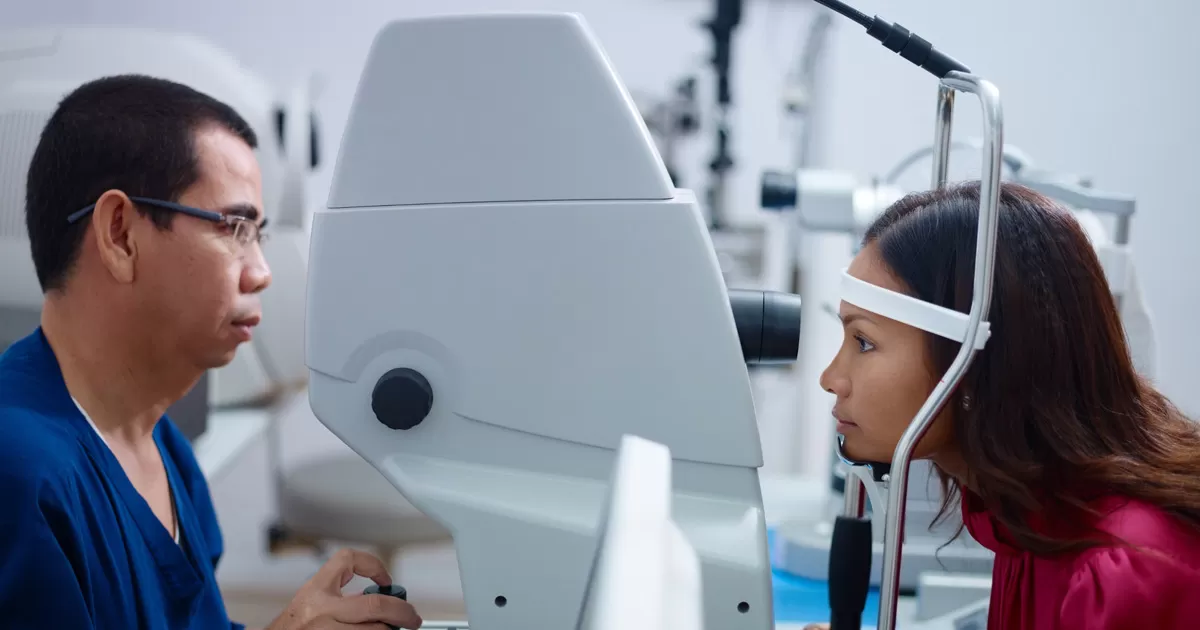
- Intraocular pressure measurement (Tonometry):
- Schiotz method — a device placed on the eye surface after numbing drops; patient lies flat
- Applanation method — uses a slit lamp microscope
- Non-contact method — a puff of air is blown onto the eye; light reflection calculates pressure
- Optic nerve and retinal examination (Ophthalmoscopy) — assessing the shape and condition of the optic nerve
- Visual field testing (Perimetry) — patient focuses straight ahead while a light is moved in from the sides to map peripheral vision
- Drainage angle examination (Gonioscopy) — a special lens placed on the eye after numbing drops allows the doctor to examine the drainage angle and determine glaucoma type
- Computerized optic nerve analysis — photographs and measures the width, length, and depth of the optic nerve for detailed monitoring
Source: BangkokHealth.com